

Special Offers
100% Performance Guaranteed
Key Specifications Table
| Species Reactivity | Key Applications | Host | Format | Antibody Type |
|---|---|---|---|---|
| H | FC, ICC, IF, IHC | R | Purified | Monoclonal Antibody |
| Description | |
|---|---|
| Catalogue Number | MAB4303-I |
| Replaces | MAB4303 |
| Description | Anti-Stage-Specific Embryonic Antigen-3 Antibody, clone MC-631 |
| Alternate Names |
|
| Background Information | Stage-specific embryonic antigen 3 (SSEA-3) is a glycosphingolipid composed of five carbohydrate units connected to a sphingolipid. Sphingolipids are key players in cell signaling and the SSEA-3 is a well known marker of stem cells (ESCs, MSCs, iPSCs, CSCs) differentiation, where SSEA-3 rapidly disappears when the cells start to differentiate. |
| Product Information | |
|---|---|
| Format | Purified |
| Presentation | Purified rat monoclonal IgMκ antibody in PBS with 0.05% sodium azide. |
| Quality Level | MQ100 |
| Applications | |
|---|---|
| Application | This Anti-Stage-Specific Embryonic Antigen-3 Antibody, clone MC-631 is validated for use in Flow Cytometry, Immunocytochemistry, Immunofluorescence and Immunohistochemistry for the detection of Stage-Specific Embryonic Antigen-3. |
| Key Applications |
|
| Application Notes | Immunocytochemistry Analysis: 10µg/mL from a representative lot detected Stage-Specific Embryonic Antigen-3 in H9 cells. Immunocytochemistry Analysis: A representative lot detected SSEA-3 immunoreactivity among T cell-derived iPSCs (TiPSCs) by fluorescent immunocytochemistry (Kishino, Y., et al. (2014). PLoS One. 9(5):e97397). Immunocytochemistry Analysis: A representative lot detected the presence of SSEA-3-positive cancer stem cells (CSCs) among cultured HCT116 colorectal cancer (CRC) cells by fluorescent immunocytochemistry (Suzuki, Y., et al. (2013). Int. J. Oncol. 42(1):161-167). Immunocytochemistry Analysis: A representative lot detected SSEA-3 immunoreactivity among induced pluripotent stem (iPS) cells from human molars-derived mesenchymal stromal cells (MSCs) by fluorescent immunocytochemistry (Oda, Y., et al. (2010). J. Biol. Chem. 285(38):29270-29278). Flow Cytometry Analysis: A representative lot detected the presence of SSEA-3-positive cancer stem cells (CSCs) in five cultured colorectal cancer (CRC) cell lines, HCT116, Caco-2, DLD-1, HT-29, and SW480 (Suzuki, Y., et al. (2013). Int. J. Oncol. 42(1):161-167). Immunofluorescence Analysis: A representative lot detected a mall number of SSEA-3-positive stromal cells in normal colorectal epithelia and SSEA-3-positive cancers in colorectal adenocarcinomas by fluorescent immunohistochemistry using frozen tissue sections (Suzuki, Y., et al. (2013). Int. J. Oncol. 42(1):161-167). |
| Biological Information | |
|---|---|
| Immunogen | 4-8 cell stage mouse embryos. |
| Clone | MC-631 |
| Concentration | Please refer to lot specific datasheet. |
| Host | Rat |
| Specificity | Reacts with the Stage-specific embryonic antigen-3 (SSEA-3) that is expressed on the surface of human teratocarcinoma stem cells (EC), human embryonic germ cells (EG) and human embryonic stem cells (ES). No immunoreactivity is evident with undifferentiated murine EC, ES and EG cells. Expression of SSEA-3 is down regulated following differentiation of human EC cells. In contrast, the differentiation of murine EC and ES cells may be accompanied by an increase in SSEA-3 expression. |
| Isotype | IgMκ |
| Species Reactivity |
|
| Species Reactivity Note | Human. Predicted to react with Mouse. |
| Antibody Type | Monoclonal Antibody |
| Product Usage Statements | |
|---|---|
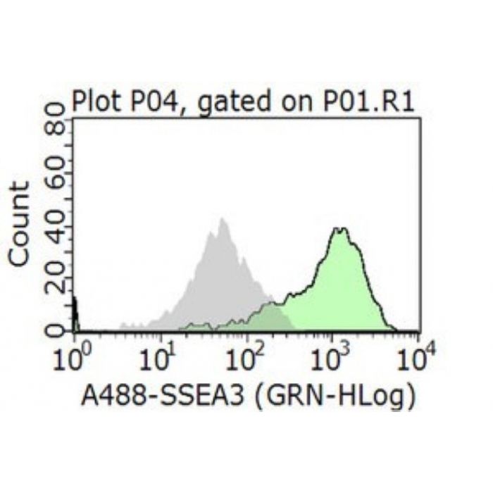
| Quality Assurance | Evaluated by Flow Cytometry in H9 cells. Flow Cytometry Analysis: 1.0 µg of this antibody detected Stage-Specific Embryonic Antigen-3 in H9 cells. |
| Usage Statement |
|
| Storage and Shipping Information | |
|---|---|
| Storage Conditions | Stable for 1 year at 2-8°C from date of receipt. |
| Packaging Information | |
|---|---|
| Material Size | 100 μg |