We use cookies to make your experience better. To comply with the new e-Privacy directive, we need to ask for your consent to set the cookies. Learn more.
Share this :
Print
Cell Signaling Neurofascin 186 (D6g6o) Rabbit mAb
Cell Signaling Neurofascin 186 (D6g6o) Rabbit mAb - CSIG (Additional S&H or Hazmat Fees May Apply)
List Price
$327.00
Your Price
$327.00
HOW MUCH YOU SAVE:
0.00 %


| NETA PART: | CSIG-15034S |
| MFG.PART: | 15034S |
| UNSPSC: | 12352200 |
| Manufacturer: | Cell Signaling |
| Size | 100 µl |
| Reactivity | H M R |
| Sensitivity | Endogenous |
| Molecular Weight (kDa) | 200 |
| Source/Isotype | Rabbit IgG |
| Application/Dilution | {Western Blotting: 1:1000, Immunoprecipitation: 1:50, Immunofluorescence (Frozen): 1:1600} |
| Storage | Supplied in 10 mM sodium HEPES (pH 7.5), 150 mM NaCl, 100 µg/ml BSA, 50% glycerol and less than 0.02% sodium azide. Store at –20°C. Do not aliquot the antibody. |
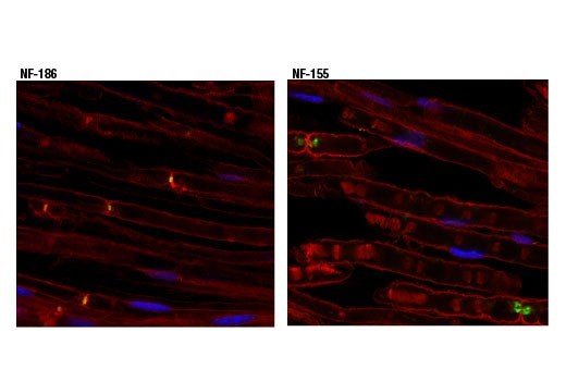
| Specificity/Sensitivity | Neurofascin 186 (D6G6O) Rabbit mAb recognizes endogenous levels of total neurofascin 186 protein. |
| Species Reactivity | Human, Mouse, Rat |
| Source/Purification | Monoclonal antibody is produced by immunizing animals with a synthetic peptide corresponding to residues surrounding Thr1108 of human neurofascin 186 protein. |
| Background | Myelinated axons contain un-myelinated gaps called nodes of Ranvier. These regularly spaced gaps are critical for the proper propagation and rapid conduction of nerve impulses in the central and peripheral nervous system (1). The structure and organization of the nodes of Ranvier is dictated by interaction between the axon and glial cells (2). Voltage-gated sodium channels concentrated at the nodes and potassium channels clustered at the paranodes are responsible for propagation of the action potentials (3,4). Other proteins that contribute to the architecture and function of the nodes of Ranvier include βIV spectrin (5), ankyrin-G (6), and the L1 cell adhesion molecules, neurofascin and NrCAM (7,8).Alternative splicing produces several neurofascin isoforms that differ in temporal and spatial expression. Neurofascin 186 is expressed in axons where it is concentrated at the nodes. Research studies indicate that neurofascin 186 is responsible for nodal assembly and clustering of sodium channels (9). Neurofascin 155 is expressed in glial cells and is localized to myelin paranodes. Interactions between neurofascin 155 and the contactin-associated protein (Caspr) tether the myelin sheath to the axon (10). N-linked glycosylation results in two forms of neurofascin 155 (high and low) that are differentially expressed during development (11). |
| SKU | CSIG-15034S |
|---|---|
| Supplier Part Number | 15034S |
| UM | UNIT |
| UNSPSC | 12352200 |
| Manufacturer Name | Cell Signaling |
| MSDS URL | https://www.cellsignal.com/contents/technical/safety-data-sheet-(sds)/resources-safety-data-sheets |
| Temperature | -20C |
| ProductLine | CSIG |
| Qty | 1 |
| MinOrderQty | 1 |
| Weight | 7.00 |
| Lead Time | 5 Business Days |