

Special Offers
100% Performance Guaranteed
Key Specifications Table
| Species Reactivity | Key Applications | Host | Format | Antibody Type |
|---|---|---|---|---|
| H | ICC, IHC, IP, WB | M | Culture Supernatant | Monoclonal Antibody |
| Description | |
|---|---|
| Catalogue Number | 05-915-25UL |
| Brand Family | Upstate |
| Trade Name |
|
| Description | Anti-N-Cadherin Antibody, clone 13A9 |
| Alternate Names |
|
| Background Information | Cadherin-2 (UniProt P19022; also known as CD325, CDw325, N-cadherin, Neural cadherin) is encoded by the CDH2 (also known as CDHN, NCAD) gene (Gene ID 1000) in human. Cadherins constitute a family of calcium-dependent cell-cell adhesion proteins that play important roles in the embryonic development and maintenance of normal tissue architecture. Cadherins are composed of an extracellular domain (a.a. 160-724 of human N-cadherin) with five homologous repeats that mediates adhesion, a single pass transmembrane domain (a.a. 725-745 of human N-cadherin), and a conserved cytoplasmic domain (a.a. 746-906 of human N-cadherin) that interacts with catenins to link cadherins to the actin cytoskeleton. In addition, a known Src substrate p120ctn also modulate the strength of cadherin-dependent adhesion by interacting with cadherins at their intracellular juxtamembrane domain. Cadherins are synthesized as precursor proteins that must be proteolytically cleaved to generate functional, mature proteins. Newly synthesized proN-cadherin (a.a. 1-906) is phosphorylated and proteolytically processed prior to transport to the plasma membrane. In addition, Plakoglobin (gamma-catenin) and beta-catenin associate only with phosphorylated proN-cadherin, whereas p120ctn can associate with both phosphorylated and non-phosphorylated proN-cadherin. The N-terminal signal and propeptide (a.a. 1-25 and 26-159 of of human N-cadherin) region is proteolytically removed and a core N-cadherin-catenin complex is assembled in the endoplasmic reticulum or Golgi compartment prior to localization at the plasma membrane where linkage to the actin cytoskeleton can be established. |
| Product Information | |
|---|---|
| Format | Culture Supernatant |
| Control |
|
| Presentation | Mouse monoclonal immunoglobulin hybridoma culture supernatant containing 0.05% sodium azide before the addition of glycerol to 30%. |
| Quality Level | MQ100 |
| Applications | |
|---|---|
| Application | Detect N-cadherin using this Anti-N-Cadherin Antibody, clone 13A9 validated for use in Immunocytochemistry, Immunohistochemistry, Immunoprecipitation, and Western Blotting. |
| Key Applications |
|
| Application Notes | Immunohistochemistry Analysis: A representative lot immunostained the extracellular matrix of the stable plaques and in the fibrous cap region rich in vascular smooth muscle cells (VSMCs) using patients-derived paraffin-embedded internal carotid artery tissue sections (Musumeci, G., et al. (2014). Histol. Histopathol. 29(6):707-719). Immunohistochemistry Analysis: A representative lot detected strong N-cadherin immunoreactivity in paraffin-embedded rectal cancer (RC) tissues with positive regional lymph node metastasis (RLNM) status, while only weak N-cadherin immunoreactivity was detected in RC with negative RLNM, and no N-cadherin staining was seen in normal colorectal epithelium (Fan, X.J., et al. (2012). Br. J. Cancer. 106(11):1735-1741). Immunohistochemistry Analysis: A representative lot detected N-cadherin immunoreactivity in formalin-fixed, paraffin-embedded hepatocellular carcinoma (HCC) tissue sections. A significant inverse correlation was found between RUNX3 and N-cadherin expression levels (Tanaka, S., et al. (2012). Int. J. Cancer. 131(11):2537-2546). Western Blotting Analysis: A representative lot detected an upregulated N-cadherin expression in CCL185 carcinoma cells following transient Epstein-Barr virus (EBV) infection. The EMT-like phenotype remained even after viral loss by culture selection pressure withdrawal (Queen, K.J., et al. (2013). Int. J. Cancer. 132(9):2076-2086). Western Blotting Analysis: A representative lot detected N-cadherin in Hep3B, Huh7, HLF and SK-Hep1 human hepatocellular carcinoma (HCC) cell lysates (Tanaka, S., et al. (2012). Int. J. Cancer. 131(11):2537-2546). Western Blotting Analysis: A representative lot detected both the unprocessed (pro-) and processed (mature) forms of N-cadherin in HeLa cell lysate (Wahl, J.K. 3rd., et al. (2003). J. Biol. Chem. 278(19):17269-17276). Western Blotting Analysis: A representative lot detected N-cadherin in WI-38 human fibroblast lysate, but not in JAr human placental choriocarcinoma cell lysate (Knudsen, K.A., et al. (1995). J. Cell Biol. 130(1):67-77). Immunocytochemistry Analysis: A representative lot detected N-cadherin immunoreactivity localized primarily at the cell-cell borders by fluorescent immunocytochemistry staining of 1% paraformaldehyde-fixed, methanol-permeabilized HeLa cells (Wahl, J.K. 3rd., et al. (2003). J. Biol. Chem. 278(19):17269-17276). Immunocytochemistry Analysis: A representative lot detected N-cadherin immunoreactivity colocalized with those of alpha- and beta-catenin by dual fluorescent immunocytochemistry staining of fixed WI-38 human fibroblasts (Knudsen, K.A., et al. (1995). J. Cell Biol. 130(1):67-77). Immunoprecipitation Analysis: Representative lots co-immunoprecipitated alpha-catenin, beta-catenin, and plakoglobin with N-cadherin from WI-38 human fibroblast and HeLa cell lysates (Wahl, J.K. 3rd., et al. (2003). J. Biol. Chem. 278(19):17269-17276; Knudsen, K.A., et al. (1995). J. Cell Biol. 130(1):67-77). |
| Biological Information | |
|---|---|
| Immunogen | Bacterially expressed human N-cadherin cytoplasmic domain MBP fusion protein (Knudsen, K.A., et al. (1995). J. Cell Biol. 130(1):67-77). |
| Epitope | Cytoplasmic domain. |
| Clone | 13A9 |
| Concentration | Please refer to lot specific datasheet. |
| Host | Mouse |
| Specificity | Clone 13A9 recognizes N-cadherin, but not P-, E-, or M-cadherin (Knudsen, K.A., et al. (1995). J. Cell Biol. 130(1):67-77). |
| Species Reactivity |
|
| Antibody Type | Monoclonal Antibody |
| Entrez Gene Number |
|
| Gene Symbol |
|
| Purification Method | Unpurified |
| UniProt Number |
|
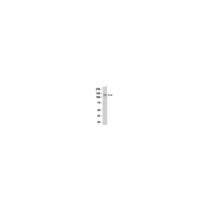
| Molecular Weight | ~140 kDa observed. Target band size appears larger than the calculated molecular weights of 82.03 kDa (mature) and 99.81/97.04 kDa (isoform 1/2 pro-form) due to posttranslational glycosylation and phosphorylation. |
| Product Usage Statements | |
|---|---|
| Quality Assurance | Evaluated by Western Blotting in HeLa cell lysate. Western Blotting Analysis: A 1:1000-5000 dilution of this hybridoma culture supernatant detected N-cadherin in HeLa cell lysate. |
| Usage Statement |
|
| Storage and Shipping Information | |
|---|---|
| Storage Conditions | Maintain for 2 years at -20°C from date of shipment. Aliquot to avoid repeated freezing and thawing. For maximum recovery of product, centrifuge the original vial after thawing and prior to removing the cap. |
| Packaging Information | |
|---|---|
| Material Size | 25 µL |