The store will not work correctly when cookies are disabled.
We use cookies to make your experience better.To comply with the new e-Privacy directive, we need to ask for your consent to set the cookies. Learn more.
Millipore Detect Neurofascin Using This Mouse Monoclonal Antibody, Anti-Pan-Neurofascin Antibody, Clone L11a/41 Validated For Use In Western Blotting & Ihc. - Mill (Additional S&H or Hazmat Fees May Apply)
Neurofascin is a ankyrin binding protein that may be involved in myelination, neurite extension, synaptogenesis, axonal guidance and neuron-glial cell interactions. There are two known isoforms, NF155 and NF186. NF155 is found in oligodendrocytes, and is involved in neurite outgrowth and neuronal adhesion. NF186 is expressed in neurons, and may optimize communication between neurons by anchoring the axon initial segment (AIS).
Product Information
Format
Purified
Control
Rat brain tissue lysate
Presentation
Purified mouse monoclonal IgG1κ in buffer containing 0.1 M Tris-Glycine (pH 7.4), 150 mM NaCl with 0.05% sodium azide.
Quality Level
MQ100
Applications
Application
Detect Neurofascin using this mouse monoclonal antibody, Anti-Pan-Neurofascin Antibody, clone L11A/41 validated for use in western blotting & IHC.
Key Applications
Western Blotting
Immunohistochemistry
Application Notes
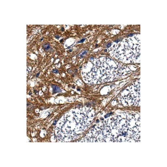
Immunohistochemistry Analysis: A 1:50 dilution from a representative lot detected Pan-Neuorfascin in rat pons tissue.
Biological Information
Immunogen
Recombinant protein corresponding to rat Pan-Neurofascin.
Clone
L11A/41
Concentration
Please refer to the Certificate of Analysis for the lot-specific concentration.
Host
Mouse
Isotype
IgG1κ
Species Reactivity
Rat
Species Reactivity Note
Demonstrated to react with Rat. Other Homologies: Human (99% sequence homology).
Antibody Type
Monoclonal Antibody
Entrez Gene Number
NP_001153785
Gene Symbol
Nfasc
Purification Method
Protein G Purified
UniProt Number
P97685
Molecular Weight
~185 kDa and ~155 kDa observed. Three isoforms are known to exist at 138 kDa, 134 kDa, and 132 kDa due to alternative splicing. This protein has been observed at ~186 kDa, ~180 kDa, ~166 kDa and ~155 kDa (Kriebel, M., et al. (2012). Int J Biochem Cell Biol. 44(5):694-697.). This protein may undergo post-translational modification.
Product Usage Statements
Quality Assurance
Evaluated by Western Blotting in rat brain tissue lysate.
Western Blotting Analysis: 1 µg/mL of this antibody detected Pan-Neurofascin in 10 µg of rat brain tissue lysate.
Usage Statement
Unless otherwise stated in our catalog or other company documentation accompanying the product(s), our products are intended for research use only and are not to be used for any other purpose, which includes but is not limited to, unauthorized commercial uses, in vitro diagnostic uses, ex vivo or in vivo therapeutic uses or any type of consumption or application to humans or animals.